Bengaluru: Researchers at the Indian Institute of Science (IISc) and their collaborators have unravelled the real-time mechanism involved in biological processes like cell division, cell mobility, transportation of nutrients into cells, and viral infections.
Although it is known that seamless transition of cell membranes between distinct 3D configurations is responsible for these processes, the experiment conducted by the IISc team throws light on the actual mechanism involved in the process.
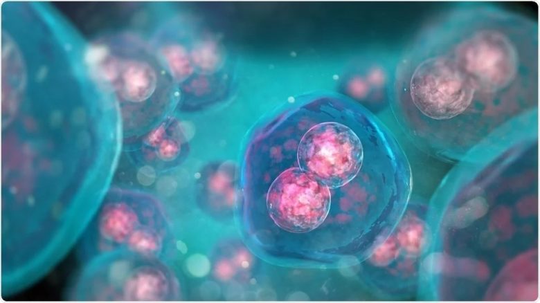
The researchers studied colloidal membranes, which are micrometre-thick layers of aligned, rod-like particles as they exhibit many of the same properties as cell membranes.
Unlike a plastic sheet, where all the molecules are immobile, cell membranes are fluidic sheets in which each component is free to diffuse.
“This is a key property of cell membranes which is available in our [colloidal membrane] system as well,” said Prerna Sharma, associate professor at the Department of Physics, IISc, and corresponding author of the study published in the journal ‘Proceedings of the National Academy of Sciences’.
The colloidal membranes were composed by preparing a solution of rod-shaped viruses of two different lengths: 1.2 micrometre and 0.88 micrometre.
The researchers studied how the shape of the colloidal membranes changes as one increases the fraction of short rods in the solution.
“I made multiple samples by mixing different volumes of the two viruses and then observed them under a microscope,” said Ayantika Khanra, a PhD student in the Department of Physics and the first author of the paper.
The researchers observed that when the saddles merged laterally, they formed a bigger saddle of the same or higher order.
However, when they merged at an almost right angle, away from their edges, the final configuration was a catenoid-like shape. The catenoids then merged with other saddles, giving rise to increasingly complex structures, like trinoids and four-noids.
A key insight of the study was to show that the Gaussian curvature modulus of the membranes increases when the fraction of short rods is increased. This explains why adding more short rods drove the membranes towards saddle-like shapes, which are lower in energy. It also explains another observation from their experiment where low-order membranes were small in size, while high-order membranes were large.
(IANS)


